Our Services
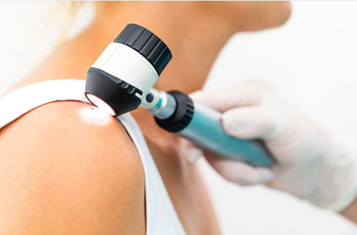
Our Adelaide based skin cancer clinic is renowned for its hi-tech skin cancer checks. During the skin cancer check, you will be asked to strip down to your underwear, and our expert doctors will undertake a thorough visual analysis of the skin and scan your whole body using the MoleMax HD machine. If any areas look a little suspicious, they will be fully analysed up close using the ultra-high magnification of the MoleMax HD which allows us to see the structure of the skin.
If something needs further investigation, then the skin doctor will talk you through the options which may involve taking a small biopsy
of the skin and sending it to the lab for further analysis or they may recommend a treatment for you. We then keep your MoleMax HD body map on file, so we can use it to map any changes in your skin at future check-ups.
Skin cancer checks are painless, completely
safe and usually last around 30 minutes
We ask that you refrain from wearing makeup, fake tan or nail polish on the day of the appointment.
As part of our commitment to early detection and prevention of skin cancer, your Skin Matters doctor will also talk to you about how to examine yourself on a regular basis between check-ups and what to look out for.
In general, we would recommend that people over the age of 50, those with a family history of melanoma should have a skin cancer check every year. We would also recommend that people under the age of 50, with no family history, should have skin cancer check every two years.
Skin Cancer Clinic
in Adelaide’s East
All of our skin doctors are trained to conduct minor skin cancer treatments or remove benign (non-cancerous) moles, benign lesions or skin tags within the Adelaide CBD clinic location. However, if you do need to see a specialist for further treatment, we can immediately refer you to the relevant skin cancer specialists on site or any other specialist as required.
Your Skin doctor will discuss the best treatment options to get you the best possible outcomes for any skin cancer or skin issue that may be discovered. The treatments available from our Skin Doctors include biopsies, excisions, and cryotherapy.


Skin Lesion Biopsy
A biopsy is a simple procedure that is used for diagnostic purposes only. A local anesthetic is applied to numb the area, and a small sample of skin will be taken to be sent off to the lab for further analysis.
A biopsy can be collected in different ways, the two most popular methods being a punch biopsy or shave biopsy.
A punch biopsy is performed using a circular-shaped instrument called a punch. The punch is placed directly over the lesion, pushed down, then slowly rotated to remove a small, round piece of skin.
Alternatively, a shave biopsy requires the use of a scalpel (a sharp surgical knife), to shave a section of the lesion off.
In both cases, the biopsy site is then covered by a sterile dressing or bandage, and because the cut size is small, sutures (stitches) are rarely required. Please be aware that there is always a possibility of soreness and scarring, although our highly skilled doctors always try to minimise the visual impact of any scars.
Skin Lesion Excisions
Certain skin cancers may only require a simple excision, where our highly experienced Skin Doctors will simply remove the cancerous growth, tumor, skin lesion or mole. The treatment will be done on site, under a local anesthetic and the method of excision is very similar to that of a biopsy
However, when removing an entire lesion, mole or growth, the cut may be deeper and larger, and is more likely to require the use of either standard or dissolving sutures (stitches).


Skin Mapping
Using our state of the art, MoleMax HD system camera, we are able to take high quality photos to help with the early detection of Melanoma and other cancerous growth, tumour, skin lesion or mole. The MoleMax HD system allows for accurate photos of the skin without the need of additional oils or gels.
This procedure involves multiple photos across a period of time to monitor changes of a certain area and could require later surgical removal.
Cryotherapy Treatment
Cryotherapy is a relatively quick and simple procedure which is highly effective on ‘pre-cancerous skin cells’ called Actinic Keratoses (or Solar Keratosis), certain types of skin cancers and resistant warts.
This technique uses liquid nitrogen, which is applied directly to the lesion for a short, but specific time – usually just a few seconds and is used to freeze and kill off abnormal cells. A blister may develop within hours of the treatment which is expected to heal within 7 – 14 days.
